• Angiogram/Cardiac Catheterization


    3/21/2015

    An angiogram is a diagnostic procedure that provides detailed x-ray pictures of your heart and its blood vessels.

    An angiogram will show:

    • That blood flow to your heart is not being restricted by blockages -- a finding that lets you and your doctor know your symptoms are not related to your heart; or
    • That the arteries to your heart are narrowed or blocked, exactly where the blockages are located and their size -- information that will enable the cardiologist to develop a treatment plan.

    How Does an Angiogram Work?

    Your cardiologist performs an angiogram to "see" any blockages in the heart (coronary) arteries, the vessels through which blood flows to your heart muscle. To get this special view, a catheter (a small, flexible tube) is inserted into a large vessel in your upper thigh or arm, and then guided through the arteries to the heart. A special “contrast” dye is injected into the catheter and to the coronary arteries and heart.

    The dye makes it possible for an x-ray camera to record a “movie” of your heart and its arteries. The movie, which you and your doctor may view on the TV screen, shows blood flow through the arteries of the heart and the location of any blockages. The angiogram also provides details about the size and shape of any blockages. This information is critical because it helps the cardiologist plan the best approach for treating each blockage.

    How Is an Angiogram Performed?

    Before the Angiogram

    Your cardiologist or nurse practitioner will offer instructions on how to prepare for the angiogram.

    • If you take medications, such as blood-thinners, consult with your cardiologist about whether you should stop taking them before the angiogram. Aspirin should be continued.
    • You will be asked not to eat or drink anything for several hours before the procedure.
    • Your blood count, kidney function and blood clotting tests may be performed beforehand.
    • You will receive medications to help you relax before the procedure begins.

    Having an angiogram requires a visit to a hospital to a special room called the catheterization laboratory, or “cath lab.” The cath lab is equipped with an x-ray camera and a TV monitor (screen) on which the cardiologist views your heart and arteries.

    During the Angiogram

    This content requires Flash Player.

    In this video, Dr. Mark A. Turco performs an angiogram on a woman with a family history of heart disease.

    An angiogram typically takes from 45 minutes to one hour. As the procedure begins, a nurse inserts an IV (intravenous line) into a vein in your arm. The IV allows you to receive fluids and medications easily. If you become anxious during the angiogram, you will receive more medications to help you relax. 

    During the angiogram as you lie on the table, you are mildly sedated but awake throughout the procedure. A specially trained cardiologist performs the procedure in an area called a catheterization laboratory, or “cath lab,” that is equipped specifically for the procedure.

    After you are relaxed, the doctor will use a small needle to inject lidocaine, a local anesthetic, to numb an area in the groin, or upper leg, in the arm or wrist. This needle prick could be the only pain you will feel throughout the procedure. The procedure is typically painless.

    The femoral artery in the groin - near where your leg bends from the hip - is one of the blood vessels doctors most commonly use to insert a catheter (a flexible tube that is smaller than the vessels) and thread it through the arteries to the heart to perform the angiogram. Instead of the femoral artery, your doctor may choose to insert the catheter in the brachial artery in the inside of the elbow or the radial artery in the wrist.

    From this “access” point in your leg or arm, the catheter is threaded through the arteries to your heart. Because there are no nerves in your arteries, you will not feel the catheter passing through the blood vessels.

    The x-ray camera helps the physician guide the catheter to your heart. When the catheter is properly positioned, the cardiologist injects a contrast dye (radiographic contrast agent) through the catheter into the heart and its arteries. Most people do not feel the dye injection. However, some feel minor discomfort, typically lasting only a few seconds, in their chest. A few feel lightheaded or nauseous.

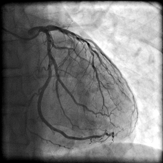
    When the x-ray beam passes through the dye, the arteries appear in black silhouette on a white background. If you have blockages, they appear as white areas. The x-ray camera records a “movie” of your heart’s pumping chamber and arteries. The movie is recorded as a medical digital image. (Not exactly ready for your home DVD player.)

    By enabling the cardiologist to see blood flow and the size, shape and length of any blockages, the angiogram provides vital information for planning the best approach to treating each one.

    Coronary AngiogramAfter the Angiogram

    If the angiogram shows serious blockages, the interventional cardiologist may immediately perform a coronary interventional procedure, such as balloon angioplasty and stenting, to open the blockage and restore blood flow to your heart. Or your doctor may refer you for coronary bypass surgery, a surgical method for restoring blood flow. If your angiogram shows plaque build-up that does not require immediate attention, your doctor will review the pictures and study your condition in more detail before recommending a plan of action, which may include lifestyle changes and/or medications.

    Care in the Hospital After the Angiogram

    When the procedure is complete, the catheter is removed and the doctor may use a device to seal the artery puncture site. Otherwise, the doctor or nurse applies pressure to the point on your leg or arm at which the catheter was inserted and holds it for about 20 minutes.

    After the wound is dressed, and if the catheter was inserted in your leg, you will be asked to lie still and avoid bending your leg or lifting your head. You may need to be still for two to six hours after the catheter is removed.

    Care at Home After the Angiogram

    After you return to your home, you may notice a bruise in the area where the doctor inserted the catheter. It is caused by blood that has escaped from the vessel under the skin. A small - and sometimes larger - bruise is normal. It is not necessary to report bruising to your doctor, even if the area is large. Usually, it clears up over a period of one to three weeks.

    You should call the doctor or nurse practitioner if you have:

    • Bleeding, drainage or painful swelling at the site where the catheter was inserted.
    • Swelling or weakness and/or numbness in the leg or arm in which the catheter was inserted.

    Is an Angiogram Safe?

    Angiograms are generally safe. However, there are risks with any test. Bleeding, infection and irregular heartbeat can occur. More serious complications, such as heart attack, stroke and death can occur, but they are uncommon.

    If you are allergic to iodine or x-ray dye, you should let your physician or nurse practitioner know so that medication to avoid an allergic reaction can be prescribed. The contrast dye typically used during the angiogram procedure contains iodine. Some patients have a minor reaction to the x-ray dye, such as a skin rash or itching. The chance of a life-threatening reaction to the dye is very small. X-ray dye very rarely causes serious or permanent kidney damage, especially if kidney function was normal before it is used. However, those who have weakened kidney function, due to diabetes or high blood pressure, may face a greater risk of further deterioration in kidney function. If it occurs, the deterioration is often temporary, but sometimes it may be permanent.

    As a procedure that uses x-ray technology, angiograms do expose patients to ionizing radiation. Safety guidelines and equipment in place in the catheterization lab are designed to limit this exposure. For more information about diagnostic testing and radiation, see the SecondsCount Guide to Radiation Safety in Medical Testing.

    Other Tests May Be Performed During the Angiogram

    During your angiogram, your interventional cardiologist may perform other tests to determine the severity of your heart condition, including the degree to which your heart arteries are narrowed by plaque buildup and whether oxygenated blood is flowing to your heart. These tests include optical coherence tomography (OCT) and intravascular ultrasound (IVUS).

    These tests can be performed while you are having your angiogram and can provide your interventional cardiologist with information to help guide treatment decisions. To learn more about these tests, click on the following links:  

    The following questions can help you talk to your physician about having an angiogram. Consider printing out or writing down these questions and taking them with you to your appointment. Taking notes can help you remember your physician’s response when you get home.

    • What information do you hope to gain from the angiogram?
    • What potential benefits may an angiogram have for me?
    • What are the alternatives to having an angiogram?
    • What risks can I expect from undergoing an angiogram?
    • Do I need to have an empty stomach before the procedure? Should I withhold any of my medications? Are there any medications that I will need to take?
    • How will you enter my body to perform the angiogram? (Common entry sites are the artery in the groin or the artery in the wrist.)
    • How much radiation will I be exposed to during the angiogram?
    • What will happen if a blockage is discovered?
    • Under what circumstances might an angiogram result in immediate angioplasty and stenting or a referral for bypass surgery?
    • Will I have limitations after the procedure? Will I need to have someone who can drive me home?