Optical Coherence Tomography
(OCT)
Overview
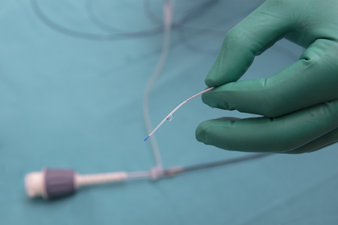
Optical coherence tomography (OCT) is a type of advanced medical imaging that uses light waves to take near-photographic quality cross-section pictures of living tissue. The test, which received regulatory approval for cardiac use in the United States in 2010, is performed as part of an angiogram.
In interventional cardiology, OCT imaging is used to take detailed images of blood vessel walls, which help to determine the extent and burden of atherosclerotic plaque (buildup of cholesterol and other material) in your arteries. This plaque can build up and cause chest pain called angina. This detailed understanding can help an interventional cardiologist determine whether a stent is needed to treat your blockage and, if so, where to best place it. After a stent has been placed, OCT can also be used to see how well the stent is covering the plaque and ensure it’s expanded to the correct size for your vessel.
More recently, studies have tested whether OCT can be used to help guide an angioplasty and stent placement, rather than being used before or after the procedure. OCT-guided procedures may offer better procedural and in-hospital outcomes.
